

Internal Structure of a Leaf
 |
| Figure: internal structure of a leaf |
When
the leaf is cut in cross-section and seen under a microscope, the below
structures are seen:
 |
| Figure: Leaf anatomy |
- Cuticle:
Cuticle is a transparent waxy layer covered
on the upper surface of the leaf
Cuticle is made up of wax which is secreted by the epidermal cells
Functions
of the cuticle:
a) Cuticle
allows light to pass through
b) Cuticle
reduces water loss (acts as a waterproofing of the leaf)
- Epidermis
Epidermis of the leaf consists of a single
layer of cells surrounding the whole leaf.
The epidermal cells of the leaf don’t contain
chloroplast.
Functions
of the epidermis:
a) Acts
as a protective layer
b) Keeps
the leaf’s shape
Epidermis of the leaf is divided into
A.
Upper
epidermis
- Upper
epidermis is found on the upper surface of the leaf and can be seen when the
leaf is cut in cross-section and observed under microscope.
- Cells
of the upper epidermis are transparent
- No
stomata are present
Functions of the upper epidermis:
a) Allow
light to pass through
b) Act
as a barrier to micro-organisms
c) Secrete
wax
B.
Lower
epidermis
- Lower
epidermis is found on the lower surface of the leaf and can be seen when the
leaf is cut in cross-section and observed under microscope.
- Stomata
are present
Functions of the lower epidermis:
a) Act
as a protective layer
b) It
is the site of gaseous exchange into and out of the leaf as stomata are present
- Mesophyll tissue
Mesophyll is the tissue between upper and
lower epidermis of the leaf
Mesophyll tissue consists of:
a) Palisade
cells (palisade mesophyll cells) ad
b) Spongy
cells (spongy mesophyll cells)
Palisade Mesophyll:
- Palisade
cells are elongated cells and box-like shape
- Palisade
cells are found below the upper epidermis
- Palisade
cells are packed together
- Palisade
cells contain maximum amount of the leaf’s chloroplasts
- Palisade
cells are the main region for photosynthesis in the leaf
- Palisade
cells receive carbon dioxide by the diffusion from air spaces in the spongy
mesophyll.
- Chloroplasts
in the palisade cells are able to move within the cytoplasm. During dim light, chloroplasts in the palisade
cells move to the upper parts of the cell allowing them maximum absorption of
light. In bright light, chloroplasts
in the palisade cells move to the lower parts of the cell for protection from
the bleaching effects of intense light.
Function of the palisade cells:
i.
Palisade cells are the photosynthetic site in
the leaf
Spongy Mesophyll:
- Spongy
cells are spherical and irregularly shaped cells
- Spongy
cells are loosely packed together
- The
air spaces between spongy cells are known as intercellular air spaces.
- Air
spaces between spongy cells allow gaseous exchange in the leaf.
- Spongy
cells allow of gaseous exchange in the leaf.
- Spongy
cells contain viewer chloroplasts
Function of the spongy cells:
i. Spongy
cells are site of gaseous exchange in the leaf.
- Vascular bundles
This is the leaf vein, made up of both xylem and phloem.
Xylem
vessels bring water and minerals to the leaf cells.
Phloem
vessels transport sugars and amino acids away from the leaf cells
(this is called translocation).
They also provide support for the leaf
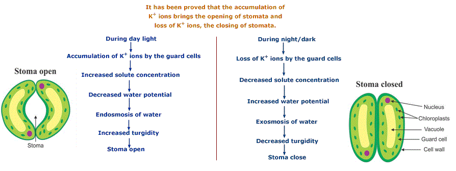
- Stomata
Stomata are tiny pores found at the lower
epidermis or underside of the leaf.
Stomata are always open during day time and
close in the night time.
Each stoma is surrounded by a pair of guard cells.
Guard cells control whether the stoma is open
or closed.
Carbon dioxide diffuses in and oxygen
diffuses out of the stomata during photosynthesis.
Water vapour passes out of the stomata during
transpiration.
Functions
of the Stomata:
I.
Allow gaseous exchange
II.
Transpiration takes place
Function
of the guard cells:
I.
Control whether the stoma is open or closed
 |
| Figure: Open and closed stoma |


0 Comments